In the realm of robotic hernia surgeries, the significance of imaging cannot be overstated—it truly stands as a cornerstone of the surgical experience! From the outset, imaging plays a vital role by providing intricate anatomical maps that aid in preoperative planning. This invaluable resource allows surgeons to strategize effectively, setting the stage for a successful operation.
As the surgery unfolds, real-time navigation facilitated by advanced imaging technologies empowers surgeons to make precise decisions on the fly, enhancing the overall safety of the procedure. Post-surgery, imaging continues to shine by monitoring the healing process, ensuring that everything is progressing as it should.
The integration of innovative tools such as 3D imaging, real-time feedback systems, and AI-driven analysis takes these advantages to a whole new level. These technological advancements not only bolster the surgeons’ capabilities but also significantly reduce risks and improve surgical outcomes.
As we continue to explore the evolving landscape of robotic hernia surgery, the transformative impact of dynamic imaging systems becomes increasingly clear. It’s an exciting journey, and the future of this field holds promising developments that will only enhance the surgical experience for patients and practitioners alike!
You might wonder why imaging is so essential in robotic hernia surgeries. Consider how it enhances preoperative planning, guides surgeons during the procedure, and helps monitor patient recovery post-surgery. In the upcoming sections, you’ll discover how each of these aspects plays an important role in achieving successful surgery outcomes.
In the field of robotic hernia surgeries, preoperative imaging plays an essential role. It’s your first step in the thorough preoperative planning process, laying the groundwork for a successful procedure. This vital stage is where you get to study, strategize, and structure your surgical approach.
Imaging provides a detailed map of the patient’s anatomy, pinpointing the exact location, size, and type of hernia. It’s like having X-ray vision, allowing you to see beneath the surface and understand the unique complexities of each case. You’re not flying blind; instead, you’re equipped with important data that can greatly influence surgical outcomes.
Robotic surgery relies heavily on this visual information. The precision of your robotic tools is only as good as the clarity of your imaging. It’s the difference between a sharp, well-defined target and a blurry, ambiguous one. By enhancing your view, imaging elevates your ability to perform accurate, minimally invasive hernia surgeries.
Charting the complex environment of a patient’s body during robotic hernia surgery is like steering a ship through uncharted waters. It’s here that imaging technology sweeps in as your guiding star.
As you navigate these internal landscapes, imaging technology provides visual clarity, guaranteeing precision in every surgical movement. Intraoperative navigation, a cornerstone in robotic surgery, hinges on this technology. It feeds real-time, high-resolution images that orient you, helping avoid critical structures while zeroing in on the hernia site.
Imagine trying to perform robotic hernia surgery blindfolded. You’d be hard-pressed to guarantee safety and efficacy without the aid of your eyes. That’s how essential imaging technology is in intraoperative navigation. It’s your eyes, granting you a virtual sightline into the patient’s body.
In the grand scheme of things, this technology not only enhances the accuracy and safety of hernia surgery but also reduces operative time. Remember, the right navigation can make all the difference between a successful voyage and a shipwreck. In your case, it’s the difference between a successful surgery and potential complications. It’s clear: imaging technology is the invaluable compass guiding your surgical journey.
Moving beyond the actual procedure, let’s now cast our gaze on the postoperative phase. Postoperative imaging plays a pivotal role in monitoring and follow-up after robotic hernia repair. It’s not just about patching the hernia and moving on. You need to monitor the patient’s progress, and that’s where imaging comes into play.
After your patient undergoes robotic surgery for hernia repair, postoperative imaging becomes your eyes. It allows you to see how the patient is healing internally. It helps you track the success of the operation and flag any complications early. You can detect issues, such as recurrence or mesh migration, that could potentially cause discomfort or lead to reoperation.
Follow-ups, coupled with imaging, form an essential part of postoperative care. They offer an opportunity to address any concerns the patient may have and reassure them about their recovery. They also allow you to adjust the treatment plan if necessary, based on the imaging findings.
You’re now stepping into the domain of imaging modalities in robotic hernia repairs. Think about the precision of CT scans, considered the gold standard in this field. Then, consider the expanding role of MRI in complex cases and the unique applications of ultrasound in robotic surgery.
In the domain of imaging modalities for robotic hernia repairs, CT scans stand as the gold standard for precision. You’ll find the clarity and detail they provide indispensable in planning and executing robotic surgery. Understanding the unique value of CT scans will enhance your mastery of this field.
Firstly, a CT scan’s high-resolution images offer a precise anatomical map. This is critical when you’re using a surgical robot to mend a hernia. You’ll be able to identify essential structures, assess the hernia’s size and location, and plan the ideal surgical route.
CT scans also provide a real-time view during surgery. This allows for adjustments on the fly, ensuring the surgical robot is always in the right place. Additionally, CT scans can identify and help avoid potential complications which could go unnoticed in less precise imaging modalities.
In short, CT scans are your best tool for maximizing the precision of robotic hernia repairs. They’re a cornerstone of medical imaging, ensuring every decision you make is informed, confident, and precise. Harness their power and see the difference they make in your surgical outcomes.
While CT scans certainly hold the fort regarding precision, let’s not forget the expanding role of MRI in complex hernia cases. As you explore deeper into the domain of robotic surgery, you’ll find that MRI serves as a critical imaging system, especially when dealing with intricate inguinal hernia cases.
MRI’s superior soft tissue contrast resolution is a game changer in accurately identifying, characterizing, and mapping hernias. This imaging modality provides an extensive, three-dimensional view of the abdominal area, aiding surgeons in preoperative planning and intraoperative navigation. It’s the difference between a tailor-made surgical procedure, and one-size-fits-all approach.
In some complex cases, the hernia may not be easily identifiable through physical examination or even a CT scan. Here, MRI’s ability to visualize hard-to-detect hernias greatly reduces the risk of missed hernias, thereby improving surgical outcomes.
Ultrasound technology, often the unsung hero in the medical field, plays a significant role in robotic hernia repairs. When integrated into a robotic platform, ultrasound provides unsurpassed visualization, enabling surgeons to steer the surgical system with precision.
The power of ultrasound lies in its ability to deliver real-time imaging. In robotic surgery, this translates to improved accuracy and safety. It’s as if you’re given a high-definition map while maneuvering through an intricate labyrinth. You can see the surgical landscape in high resolution, spot potential hazards, and plan your route accordingly.
Furthermore, with ultrasound, you’re not just seeing the surface but also what’s beneath it. You can visualize structures hidden from the naked eye, such as blood vessels and nerves. This feature is essential in hernia repairs where the goal is to fix the defect without causing collateral damage.
You’re stepping into a world where technological advancement is revolutionizing surgeries. Let’s talk about how 3D imaging and augmented reality integration, real-time imaging systems, and AI-driven imaging analysis are making robotic surgeries more successful than ever. You’ll see how these breakthroughs in imaging technology are enhancing precision, improving outcomes, and paving the way for the future of hernia repair.
Innovation’s powerful hand is reshaping the landscape of robotic hernia surgeries with the integration of 3D imaging and augmented reality. This integration enriches the perspective of surgeons, providing a real-time, three-dimensional view of the surgical field. It’s a game-changer in the domain of robotic surgery, offering unprecedented precision and control.
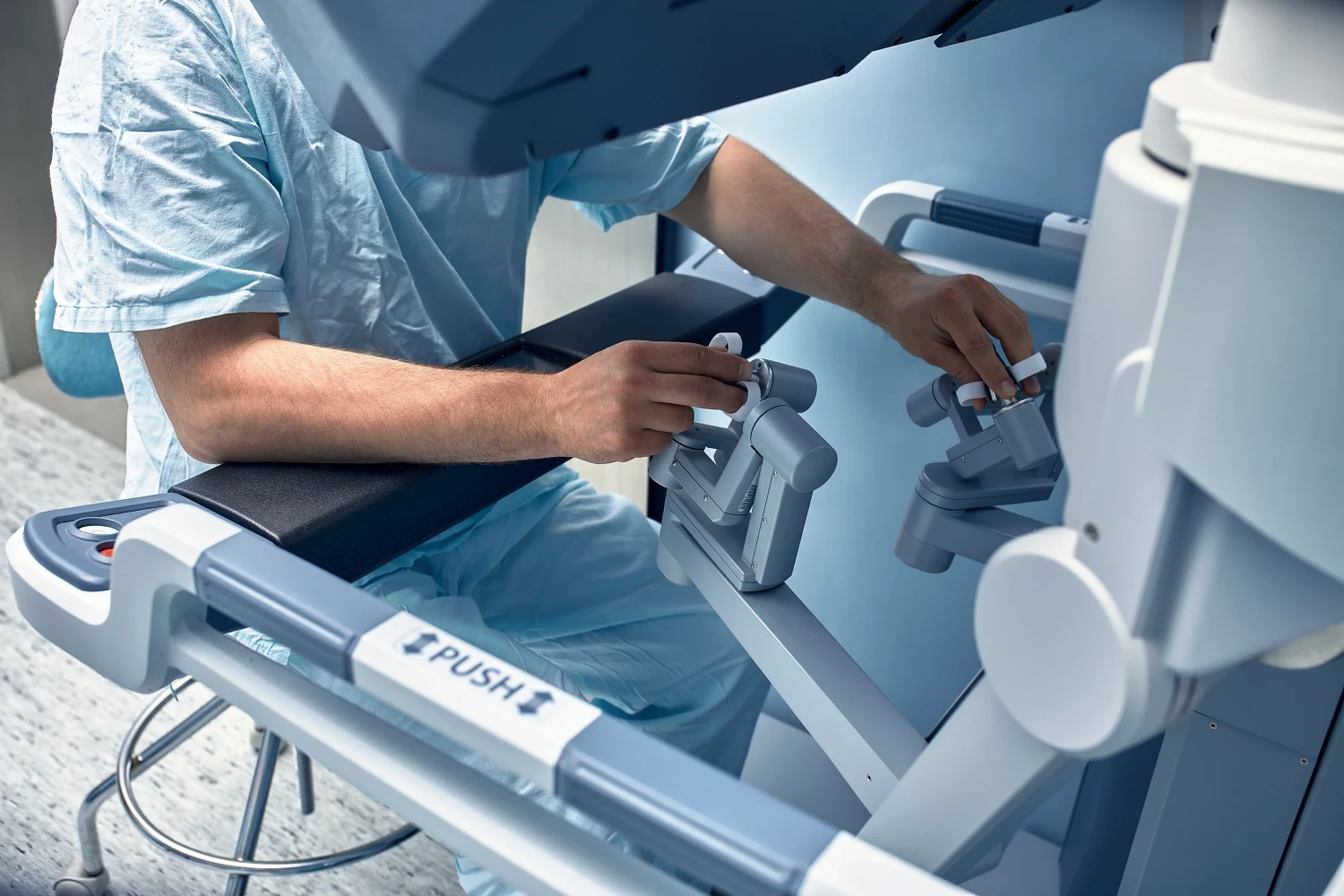
In a robotic-assisted surgery, the surgeon uses a console equipped with controls that direct robotic arms. The integration of augmented reality provides an overlay of essential data on the surgical field, enhancing surgical planning and guidance. The surgeon can see beyond the surface, getting a detailed look at the anatomy beneath, including the position of blood vessels and tissues. This information is critical for avoiding unnecessary damage and ensuring the success of the procedure.
Moreover, the 3D imaging and augmented reality integration in robotic surgery not only enhance precision but also improve the surgeon’s spatial awareness. It’s like having x-ray vision – seeing the unseen, maneuvering through the complex, and making the impossible possible. The role of imaging in robotic hernia surgeries is significant, and with the integration of augmented reality, the future of robotic surgery looks promising.
Consider the impact of real-time imaging systems, an absolute breakthrough in the domain of robotic surgery. These systems, pivotal in enhancing precision, have revolutionized how robotic hernia surgeries are performed.
Before, the lack of real-time feedback was a significant challenge in robotic surgery. Surgeons had to rely on static images taken before the procedure, which didn’t account for changes occurring during the operation. But with real-time imaging, surgeons can now view the surgical field as it is, not as it was.
Imagine you’re the surgeon. With the robotic surgical system, you’re sitting at a console, manipulating robotic arms with millimeter precision. Now, add real-time imaging to the mix. You’re not just controlling the operation, but you’re also seeing, in real time, the exact location of the hernia and the surrounding structures. You’re able to adjust your movements based on real-time feedback and perform procedures with unparalleled accuracy.
Real-time imaging hasn’t just improved the way you perform hernia surgeries, it’s also reduced complications and improved patient outcomes. It’s clear that the integration of real-time imaging systems into robotic surgical systems is one of the key imaging innovations driving the success of robotic surgery.
As a surgeon, imagine having an AI-powered imaging system as your co-pilot. Imagine the increased precision, the enhanced clarity, the amplified confidence in your decisions. AI-driven imaging analysis in robotic surgery is not a far-off dream. It’s here, and it’s revolutionizing the way you perform procedures.
This groundbreaking technology uses advanced algorithms to analyze imaging data in real time. It’s like having an extra pair of expert eyes, scanning every detail, picking up on subtleties you might miss. The robotic system, guided by this AI-driven analysis, can make pinpoint adjustments, reducing the risk of complications and improving patient outcomes.
But it’s not just about providing a clearer image. This technology also allows for better planning and execution. You can simulate various surgical scenarios, test different approaches, and choose the one that’s best for your patient. It’s about equipping you with the tools to perform at your highest level.
As you prepare for robotic hernia surgery, preoperative imaging is a critical step in your journey. It helps in identifying hernia types and severity, allowing for a customized surgical approach based on the data collected. With accurate pre-surgical mapping, risks are minimized, setting the stage for a successful procedure.
In the world of robotic hernia surgeries, preoperative imaging is a essential step. This advanced technology allows surgeons to identify the type and severity of your hernia, setting the stage for a successful operation. It’s not just about getting a good look at the problem, it’s about understanding it in depth.
Hernias come in many forms, and the severity can range from mild to severe. The type of hernia you have can determine the surgical approach needed. A hiatal hernia, for instance, requires a completely different robotic surgery approach from an inguinal hernia. Imaging can reveal this critical information before the surgery even begins.
The severity of your hernia, on the other hand, can influence the complexity of the operation. A small, uncomplicated hernia might be resolved quickly, while a large or recurring hernia could call for a more detailed surgical plan. Again, preoperative imaging plays a pivotal role in making these determinations.
In essence, preoperative imaging is your surgeon’s roadmap. It doesn’t just show them where to go; it helps them decide the best way to get there. Through imaging, robotic hernia surgery becomes a finely tuned, personalized procedure.
You might wonder how preoperative imaging fits into the big picture. Well, let’s dig in. In the world of robotic hernia surgeries, imaging data holds a prominent place. It’s not only about identifying the hernia type or severity; it’s also used to shape the surgical approach.
Using imaging data, surgeons can customize each procedure. This is essential for minimally invasive surgeries where the robot’s precision plays a pivotal role. By analyzing the images, surgeons can determine the best entry point for the robot, minimizing tissue damage and ensuring a smoother recovery.
Moreover, imaging data helps in planning the surgical pathway. It provides a roadmap for the robot to follow, avoiding critical structures, and ensuring the hernia is repaired effectively. This means your surgery isn’t a one-size-fits-all procedure. It’s customized, tailored to your unique needs.
In essence, preoperative imaging data sets the stage for success in robotic hernia surgeries. It allows for a tailored approach, making surgeries less invasive and more precise. So, while you may not see it, behind the scenes, imaging data is working hard for you.
Your surgeon’s toolkit wouldn’t be complete without accurate pre-surgical mapping. This critical step minimizes risks in minimally invasive surgery, especially when dealing with complex hernia cases.
The robotic arm, an integral component of robotic-assisted surgeries, relies heavily on precise pre-surgical maps. These maps are generated using advanced imaging technologies like CT scans and MRI. Imagine these maps as detailed blueprints that guide the robotic arm, ensuring pinpoint accuracy during the procedure.
One exciting development in preoperative imaging is the use of fluorescence imaging. This innovative technique makes body tissues glow under specific light, providing surgeons with real-time, high-resolution images. It’s like having a spotlight on the surgical field, illuminating the pathway for the robotic arm.
By accurately mapping out the surgical field beforehand, surgeons can anticipate potential issues and plan their approach accordingly. This thorough preparation reduces the likelihood of complications, optimizing patient outcomes.
The bottom line is, pre-surgical mapping is not just a nice-to-have; it’s a must-have. It’s the difference between maneuvering in the dark and having a clear roadmap, and in robotic-assisted surgery, that difference can mean everything.
Now, let’s shift our focus to the real-time guidance that intraoperative imaging provides during robotic hernia surgeries. You’ll learn how this technology enhances accuracy, improves tissue identification, and precisely guides dissections. We’ll also discuss how dynamic imaging systems help surgeons adapt to challenges that arise during these procedures.
Surgeons’ precision can be considerably enhanced with the incorporation of robotic-assisted imaging. When you’re performing hernia surgeries, accuracy is everything. And that’s where robotic-assisted surgery comes into its own, providing a clarity of vision that’s beyond the reach of the human eye.
Robotic-assisted imaging offers a unique blend of high-definition, 3D imaging that allows you to see the surgical site in exquisite detail. It’s not just about seeing better; it’s about understanding the intricacies of the human body and being able to interact with it in new ways.
With the precise imaging provided by robotic-assisted surgery, you can map the surgical site and plan your approach with a level of detail previously unimaginable. You’re not just operating on a hernia; you’re maneuvering through a highly complex system.
This kind of precision doesn’t just make surgery easier; it makes it safer. It reduces the risk of complications and improves patient outcomes. In the world of hernia surgeries, that’s not just a nice-to-have; it’s a must-have.
In achieving accuracy with robotic-assisted imaging, you’re not just a surgeon; you’re a pioneer, pushing the boundaries of what’s possible in medicine.
Intraoperative imaging serves as your GPS during surgery, offering real-time guidance that enhances tissue identification and dissection precision. When conducting robotic hernia surgeries, this tool can be your strongest ally, providing clarity in a complex landscape.
A surgical robotic system such as the da Vinci Surgical System leverages high-definition 3D visualization. With this system, you can see the surgical field in extraordinary detail, enabling you to identify and dissect tissues with remarkable accuracy. The da Vinci system’s image guidance not only increases precision but also reduces the risk of inadvertent tissue damage.
This precision is vital in robotic hernia surgeries. The surgical field is often a complex mosaic of tissues, and a slight misstep can lead to complications. With the use of intraoperative imaging, you can navigate this landscape confidently, making precise cuts and identifications that improve patient outcomes.
As you master the art of robotic hernia surgeries, you’ll often find yourself facing complex surgical situations. In these moments, dynamic imaging systems become your indispensable ally. These advanced technologies, integrated into robotic surgical platforms, offer real-time visual guidance, enhancing your ability as a surgeon to perform precise and safe operations.
Dynamic imaging systems take the guesswork out of surgery. They provide high-definition, 3D visuals, helping you to navigate through the anatomical complexities of a hernia. You can see the precise location of the hernia, its size, and proximity to essential organs. It’s like having a GPS guiding you through the body’s intricate terrain.
However, as with any technological tool, it’s vital to adapt and stay updated. Newer models of these imaging systems offer features like fluorescence imaging and infrared capabilities, pushing the boundaries of what’s visible during surgery. So, keep learning, keep mastering, and you’ll find that these imaging systems are not just tools, but partners in ensuring the best possible outcomes in robotic hernia surgeries. Remember, the key to mastery is adaptation. Harness these dynamic imaging systems, refine your skills, and conquer the challenges that lie ahead.
Now that the surgery is over, it’s your job to monitor the patient’s progress. You’ll use imaging to detect early signs of recurrence, assess healing and tissue integration, and guide any necessary post-surgery interventions. Let’s explore how you can effectively use postoperative imaging to evaluate both success and complications.
Nearly all patients who undergo robotic hernia surgeries can benefit from postoperative imaging. This essential tool serves a pivotal role in the early detection of recurrence, allowing your surgeon to swiftly intervene, if necessary.
The use of imaging in robotic hernia surgeries provides a detailed view of the surgical site, giving your surgeon invaluable insight into the healing progress. High-resolution imaging technology can reveal even the slightest aberrations, making it possible to detect hernia recurrence at the earliest stages.
Postoperative imaging isn’t just about identifying problems though; it’s also about ensuring success. It verifies that the surgery has achieved its intended outcome, providing reassurance to both you and your surgeon. In addition, it can identify any complications, such as infections or adhesions, allowing for prompt medical attention.
In the unlikely event of a recurrence, early detection through imaging allows for a more beneficial prognosis. It enables your surgeon to take immediate corrective action, reducing the risk of further complications. So, while the chance of recurrence is minimal with robotic hernia surgeries, it’s comforting to know that advanced imaging technology is there to detect it promptly, ensuring your continued health and wellbeing.
With postoperative imaging, monitoring the healing process and tissue integration after robotic hernia surgeries becomes a straightforward task. You see, robotic technology has revolutionized the way surgeons perform laparoscopic hernia repair. It provides unprecedented precision, reducing the risk of complications and enhancing the overall surgical technique.
After surgery using this advanced technology, it’s essential to keep a watchful eye on the healing process. Postoperative imaging is our reliable guide, a window into the body that allows for close examination of the surgical site. It helps us track the healing progress, ensuring that the repair is integrating well with the surrounding tissues.
The images produced can reveal any irregularities or potential issues, such as fluid collection or signs of infection. They also allow for evaluation of the hernia repair’s success, especially regarding mesh positioning and fixation.
In the aftermath of robotic hernia surgery, imaging doesn’t just monitor healing; it can also guide post-surgery interventions. Its role is pivotal, providing insights into the surgical site that can shape subsequent treatment decisions. Think of it as a map, guiding you and your medical team through the postoperative landscape.
Combining the precision of robotic assistance with the clarity of imaging, you’re able to track progress, spot potential complications, and plan any necessary interventions. This dual-tech approach enhances the efficiency and effectiveness of post-surgery interventions.
For example, if imaging reveals a fluid collection or seroma - a common occurrence after hernia surgeries - you can determine the most appropriate intervention. Sometimes, this might involve a simple watch-and-wait approach. In other cases, drainage may be required.
Or perhaps imaging uncovers a mesh displacement. With this information, your surgeon can decide whether a corrective procedure is needed.
Imagine a future where AI and machine learning are integrated into imaging for robotic hernia surgeries. Personalized imaging techniques could provide patient-centric care that’s tailored to individual needs. This isn’t just a dream, it’s the direction we’re heading as advanced imaging tools become part of routine practice.
Could the future of imaging in robotic hernia surgeries be driven by artificial intelligence (AI) and machine learning? It’s not just plausible, it’s on the horizon. As you explore the application of robotic surgery, you’ll see AI and machine learning as emerging surgical robotic technologies that are rapidly reshaping the landscape.
These innovations aren’t merely enhancing the accuracy of imaging in robotic hernia surgeries, but they’re also increasing the predictability of surgical outcomes. AI’s data-driven algorithms can identify patterns that may be invisible to the human eye, leading to more accurate diagnoses and precision in surgical procedures. Machine learning, on the other hand, learns from each procedure, improving and refining its techniques over time.
As we look towards the future of imaging in robotic hernia surgeries, the focus isn’t solely on technology like AI and machine learning. Yes, they’re vital elements, but let’s not forget the human factor. Personalized imaging techniques hold the key to truly patient-centric care.
In laparoscopic surgery, imaging is your eyes. The sharper, more detailed the image, the greater your precision. Now, imagine this image is personalized. Tailored to each patient’s unique anatomy and the specific location of their hernia. A robotic assistant, equipped with this specialized imaging, can perform a surgery that is completely customized, minimizing risks and maximizing outcomes.
But personalized imaging isn’t just about the surgery itself. It’s also about aftercare. By comparing pre and post-operative images, you can track the patient’s recovery more accurately. You can spot potential complications early and intervene effectively.
Patient-centric care is more than a buzzword. It’s a commitment to tailor every aspect of care to the individual patient. And in the domain of hernia surgeries, personalized imaging is a game-changer, empowering you to deliver on that commitment.
The integration of advanced imaging tools into your routine practice is no longer a distant reality but a tangible possibility. With the rapid progress in technology, these tools are becoming more accessible, making your robotic hernia surgeries more precise and efficient.
Integrating these tools into your routine practice isn’t merely about acquiring new equipment. It’s about redesigning your surgical workflow to utilize these resources effectively. Advanced imaging tools can provide real-time, high-resolution images, enabling you to navigate complex anatomical structures with ease during robotic hernia surgeries. This can lead to improved surgical outcomes and patient satisfaction.
However, successful integration requires a mastery of these tools. You’ll need to understand their capabilities, limitations, and how to interpret the images they produce. Training programs, workshops, and continuous education are vital in this regard.
The future of robotic hernia surgeries hinges on the effective integration of advanced imaging tools into routine practice. It’s a challenging task, but one that promises significant rewards. Embrace this technological revolution and redefine your surgical practice for the better.
Imaging stands as a pivotal force in the evolution of robotic hernia surgeries, revolutionizing how surgeons approach preoperative planning, intraoperative precision, and postoperative care. From the integration of advanced modalities like CT scans, MRI, and ultrasound to cutting-edge innovations such as AI-driven analysis and augmented reality, imaging continues to elevate surgical outcomes and redefine patient care. As the field progresses, personalized imaging and dynamic visualization systems are poised to shape the future of robotic-assisted surgeries, making them safer, more effective, and patient-centered. With a commitment to utilizing the latest imaging advancements, Dr. Brian Harkins is proud to deliver exceptional surgical outcomes, ensuring the highest standards of care for every patient.
Laparoscopic techniques involve small incisions and the use of laparoscopic instruments, providing a minimally invasive option for many procedures. A robotic approach, such as those using the da Vinci robotic surgical system, enhances this by offering greater magnification, precision, and flexibility with robotic arms mounted on a surgery platform for optimal control.
The use of the robotic platform in procedures like robot-assisted surgery allows for precise movements using robotic instruments and high-definition visuals. This minimally invasive robotic method reduces recovery time, minimizes scarring, and provides surgeons with enhanced dexterity compared to open surgery.
Single-port surgery involves using one incision to access the surgical site, often leveraging single port orifice robotic technology. Unlike traditional robotic procedures that use 4 robotic arms, this approach reduces the number of entry points while maintaining the precision and control associated with robotically assisted methods.
The da Vinci robotic surgical system has transformed gynecologic surgery by allowing for precise and minimally invasive procedures. With robotic arms mounted for improved range of motion and magnification for detailed views of the abdomen, this system enables better outcomes for conditions that previously required open surgery.
The Food and Drug Administration ensures the safety and efficacy of robotic systems like the da Vinci robotic and senhance robotic platform. This oversight is critical for approving new technologies, such as novel robotic systems and flex robotic system innovations, that continue to advance surgical care.
In pediatric surgery, laparoscopic techniques remain a staple for minimally invasive care. However, the robotic and laparoscopic methods are often combined, with systems like the intuitive surgical platform offering enhanced precision and reduced tissue trauma, critical for delicate procedures in younger patients.
Robotic systems like the flexible robotic and da Vinci robotic surgical system are increasingly utilized in spinal surgery. These platforms provide unparalleled precision, especially in complex cases, by allowing surgeons to navigate the abdominal wall and spinal anatomy with minimal tissue disruption and greater accuracy.
Transoral robotic surgery is a groundbreaking advancement, allowing for minimally invasive access to areas like the throat and base of the tongue. Using systems such as the flex robotic system, surgeons can perform procedures that once required open surgery, reducing recovery times and improving outcomes.
In bariatric surgery, the precision of robotic instruments is essential for navigating the abdomen and performing delicate procedures. The robotic operating capabilities of platforms like the da Vinci robotic surgical system enable surgeons to make smaller incisions, resulting in faster recovery and fewer complications compared to traditional methods.
A surgery program must carefully adopt new robotic platforms like the senhance robotic platform and intuitive surgical systems for general surgeries. These systems enhance outcomes by offering advanced robotic and laparoscopic capabilities, reducing surgical table time and improving surgeon ergonomics during complex procedures.
Dr. Brian Harkins is a renowned surgeon specializing in advanced, minimally invasive, and robotic surgical techniques. With a dedication to innovation and personalized patient care, he has transformed countless lives by delivering exceptional outcomes.
I want a website like this, where do i start?